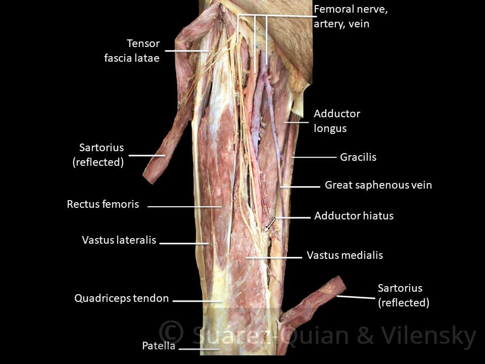
Home » Uncategories » Upper Leg Tendon Anatomy - Hip And Thigh Muscles Anatomy And Functions Kenhub / Rectus femoris these four muscles come together to form a single tendon, which inserts into the patella, or kneecap.
Sunday, 27 June 2021
Upper Leg Tendon Anatomy - Hip And Thigh Muscles Anatomy And Functions Kenhub / Rectus femoris these four muscles come together to form a single tendon, which inserts into the patella, or kneecap.
Upper Leg Tendon Anatomy - Hip And Thigh Muscles Anatomy And Functions Kenhub / Rectus femoris these four muscles come together to form a single tendon, which inserts into the patella, or kneecap.. Anatomy of the human body. Upper leg tendon anatomy : Squeeze your knees together and boom, you're contracting the adductors. Rectus femoris these four muscles come together to form a single tendon, which inserts into the patella, or kneecap. It begins in the thigh area and extends to the head of the fibula in the knee.
The rectus femoris is located in the center of the thigh, while the vastus medialis is in the middle of the said body part. This important tendon in the back of the calf and ankle connects the plantaris, gastrocnemius, and soleus muscles to. It's the area that runs from the hip to the knee in each leg. Tendons are cords made of tough tissue, and they work as special connector pieces between bone and muscle. These muscles run from the lower spine and pelvis, join together, then attach by a tendon to the upper thigh.
Physical Therapy Guide To Groin Strain Choosept Com from www.choosept.com Muscle recovery anatomy 12 photos of the muscle recovery anatomy anatomy of muscle recovery, muscle recovery anatomy, human muscles, anatomy of muscle recovery, muscle recovery anatomy Upper leg anatomy and function the upper leg is often called the thigh. Anatomy of the human body. This important tendon in the back of the calf and ankle stores the elastic energy needed for running, jumping, and other physical activity. The largest muscle masses in the leg are present in the thigh and the calf. These muscles run from the lower spine and pelvis, join together, then attach by a tendon to the upper thigh. One of the most important tendons in terms of mobility of the leg is the achilles tendon. The four muscles all extend the lower leg.
Rectus femoris these four muscles come together to form a single tendon, which inserts into the patella, or kneecap.
A muscle strain or tear may cause your thigh to look deformed. This long muscle flexes the knee. Squeeze your knees together and boom, you're contracting the adductors. The rectus femoris is located in the center of the thigh, while the vastus medialis is in the middle of the said body part. They bear the weight of the upper body. The largest muscle masses in the leg are present in the thigh and the calf. Anatomy of the human body. This tendon can get irritated from overuse, muscle weakness and muscle tightness. The uppermost of the medial thigh muscles is the pectineus muscle. A visit to an orthopedic surgeon may be needed to accurately diagnose and treat your condition. The calf comprises of 2 major muscles (gastrocnemius and soleus) both of which. One more example is the large muscle group of the quadriceps, located on the front of the upper leg. This important tendon in the back of the calf and ankle stores the elastic energy needed for running, jumping, and other physical activity.
This is why you have to indicate which biceps you are taking about when discussing one or other of these muscles. Notice the upper leg has a biceps muscle just like the upper arm does. Related posts of muscle anatomy upper leg.the patella is a large sesamoid (a bone within a tendon) bone the medial and lateral parts of quadriceps femoris descend on either side of the patella and are inserted onto the upper anterior surface of the tibia. Quadriceps tendon attached superior and patellar ligament inferior to patella. This important tendon in the back of the calf and ankle stores the elastic energy needed for running, jumping, and other physical activity.
Upper Leg Muscles And Thorax from image.slidesharecdn.com Thigh pain that comes on suddenly and limits your ability to walk could be due to a pinched nerve in your back. Anatomy of the human body. One more example is the large muscle group of the quadriceps, located on the front of the upper leg. The four muscles all extend the lower leg. Upper leg anatomy and function the upper leg is often called the thigh. Notice the upper leg has a biceps muscle just like the upper arm does. This important tendon in the back of the calf and ankle stores the elastic energy needed for running, jumping, and other physical activity. •medial thigh muscles•adductor longus muscle•adductor magnus muscle•adductor.
The quadriceps tendon works with the muscles in the front of your thigh to straighten your leg.
Tendons are strong cords of fibrous tissue that attach muscles to bones. One more example is the large muscle group of the quadriceps, located on the front of the upper leg. The anterior femoral muscles (fig. Muscle mri can provide information that is complementary to clinical, histologic, genetic, and laboratory findings for the diagnosis of neuromuscular disease. A large tear of the quadriceps tendon is a disabling injury. The hamstring portion of the adductor magnus has a similar action to these muscles, but is located in the medial thigh. The knee joint is the junction of the thigh and leg. The upper leg is composed of the femur the hamstring tendon is also connected to the tibia, immediately below the rear of the knee joint. It's the area that runs from the hip to the knee in each leg. One of the most important tendons in terms of mobility of the leg is the achilles tendon. Thigh pain that comes on suddenly and limits your ability to walk could be due to a pinched nerve in your back. Squeeze your knees together and boom, you're contracting the adductors. Muscle anatomy of upper thigh.
Muscle anatomy of upper thigh. This long muscle flexes the knee. In addition to these, the end of the iliopsoas muscle passes into the anterior. It's the area that runs from the hip to the knee in each leg. The upper leg is composed of the femur the hamstring tendon is also connected to the tibia, immediately below the rear of the knee joint.
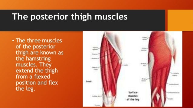
Muscles Of The Anterior Thigh Quadriceps Teachmeanatomy from teachmeanatomy.info One of the most important tendons in terms of mobility of the leg is the achilles tendon. It is also visible on the medial edge of the thigh from the anterior. Upper leg anatomy and function the upper leg is often called the thigh. These muscles run from the lower spine and pelvis, join together, then attach by a tendon to the upper thigh. The largest muscle masses in the leg are present in the thigh and the calf. The knee joint is the junction of the thigh and leg. A visit to an orthopedic surgeon may be needed to accurately diagnose and treat your condition. Anterior muscles extend your legs and flex your thighs.
The uppermost of the medial thigh muscles is the pectineus muscle.
The upper leg is composed of the femur the hamstring tendon is also connected to the tibia, immediately below the rear of the knee joint. An orthopedist can diagnose this condition. Your lower leg includes three main muscles, located behind your tibia or shinbone. This is the group of muscles that you often see body builders flexing, which protrude just above the knee and take up most of the upper leg. Rectus femoris these four muscles come together to form a single tendon, which inserts into the patella, or kneecap. Your upper leg includes seven major muscles. Upper leg tendon anatomy : It serves to attach the plantaris, gastrocnemius (calf) and soleus muscles to the calcaneus (heel) bone. Anatomy of the human body. The uppermost of the medial thigh muscles is the pectineus muscle. Upper leg tendon anatomy this mri wrist coronal cross sectional anatomy tool is absolutely free to use. Squeeze your knees together and boom, you're contracting the adductors. This chart is beautifully illustrated and offers the most comprehensive look at the muscles of the human leg available.
0 Response to "Upper Leg Tendon Anatomy - Hip And Thigh Muscles Anatomy And Functions Kenhub / Rectus femoris these four muscles come together to form a single tendon, which inserts into the patella, or kneecap."

0 Response to "Upper Leg Tendon Anatomy - Hip And Thigh Muscles Anatomy And Functions Kenhub / Rectus femoris these four muscles come together to form a single tendon, which inserts into the patella, or kneecap."
Post a Comment